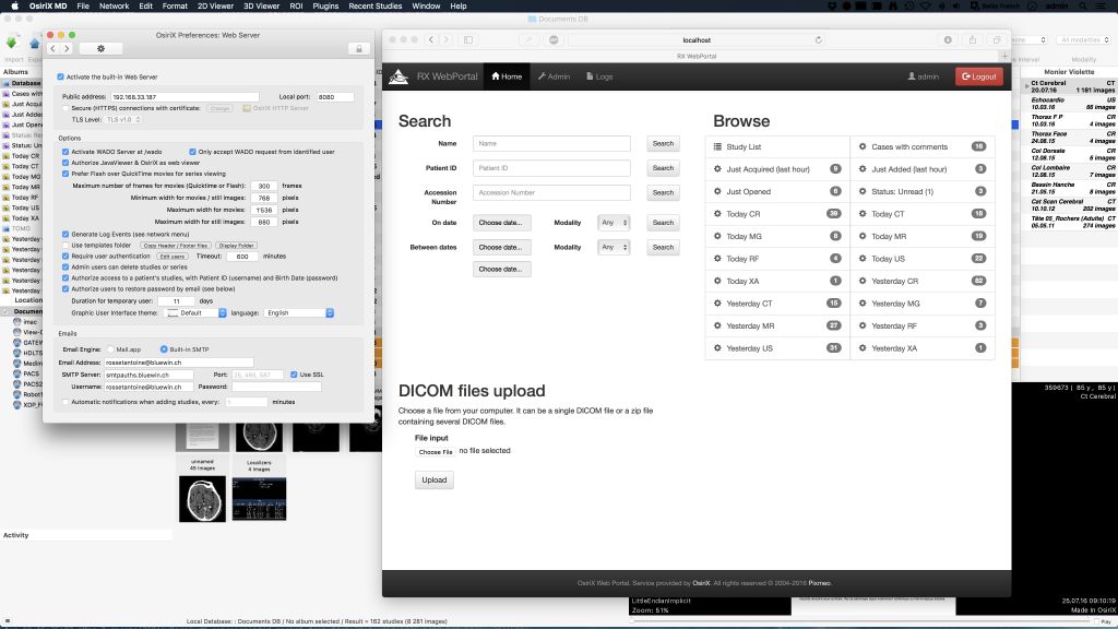
Activate Dicom Editing Osirix Dicom
We have created showing how to install RadiAnt on Mac computers with the latest macOS 10.15 update (Catalina). Apple dropped support for 32-bit applications in their latest system, so we had to prepare a special 64-bit installation package for macOS.
Check the video for more details:Disclaimer: RadiAnt DICOM Viewer is built and tested specifically for Windows platform. We do not officially support RadiAnt on platforms other than Windows.
RadiAnt can technically run on macOS systems using the open-source Wine software, however, some features may not be available or may cause program crashes. The long-awaited feature is now available in RadiAnt. It allows you to import DICOM studies from CD/DVD discs, USB flash drives, local and network folders or PACS servers, and store them on your local hard drive so that they can be easily accessed at a later date.You can also access the database to organize and quickly find studies in your collection of DICOM files.We also added a new feature to the VR module. 3D models created in RadiAnt can be, which can be opened in alternative 3D modeling software and used for 3D printing.
Patient CD/DVD DICOM ViewerDo you know how frustrating it can be to endlessly wait for a patient CD to open?Does your viewer require the installation of additional components before the images can actually be viewed?Try the RadiAnt DICOM Viewer CD/DVD! It is extremely fast, runs from CD/DVD media without installation on Windows XP SP3, Vista, Windows 7, Windows 8, Windows 8.1 and Windows 10 systems and does not have any additional software or programming requirements (e.g.NET, Java).If the user’s operating system permits, the 64-bit version is opened for better efficiency.
On older machines the 32-bit version is used. Approximately just 6MB of overhead data is added to the media.The logo image displayed after opening the application is fully customizable and can be used to show your company information to your clients. All the necessary tools close at handRadiAnt DICOM Viewer provides the following basic tools for the manipulation and measurement of images:. Fluid zooming and panning. Brightness and contrast adjustments, negative mode. Preset window settings for Computed Tomography (lung, bone, etc.). Ability to rotate (90, 180 degrees) or flip (horizontal and vertical) images.
Segment length. Mean, minimum and maximum parameter values (e.g.
Density in Hounsfield Units in Computed Tomography) within circle/ellipse and its area. Angle value (normal and Cobb angle). Pen tool for freehand drawing. Quick as lightningRadiAnt DICOM Viewer was designed to use resources as efficiently as possible.
Horos Manual
It can make use of a multiprocessor and multicore system with large amounts of gigabytes of RAM, but will also run on an old single-core machine with only 512MB RAM.A 64-bit version is provided for modern systems to keep all opened images in more than 4GB of memory, if necessary. Asynchronous reading lets you browse and process images while they are still being opened.All of this is available in one very compact application that has an installer size of just over 2MB. Search and download studies from PACS locationsThe PACS (Picture Archiving and Communication System) client feature lets RadiAnt DICOM Viewer query and retrieve studies from other PACS hosts.Supported service class users/providers are: C-ECHO SCU, C-ECHO SCP, C-FIND SCU, C-MOVE-SCU, C-STORE-SCP (Only transfers initiated from the RadiAnt DICOM Viewer are accepted. If you try to send studies from other PACS nodes without searching them first and starting the download in RadiAnt, they will be ignored).Received DICOM files are stored in a temporary folder and are deleted when RadiAnt closes. Supports multiple DICOM file typesThe software has the capability to open and display studies obtained from different imaging modalities:. Digital Radiography (CR, DX).
Mammography (MG). Computed Tomography (CT). Magnetic Resonance (MR). Positron Emission Tomography PET-CT (PT). Ultrasonography (US). Digital Angiography (XA). Gamma Camera, Nuclear Medicine (NM).
Secondary Pictures and Scanned Images (SC). Structured Reports (SR)Many types of DICOM images are supported:.
Monochromatic (e.g. CR, CT, MR) and color (e.g. US, 3D reconstructions).
Static images (e.g. CR, MG, CT) and dynamic sequences (e.g.
XA, US). Uncompressed and compressed (RLE, JPEG Lossy, JPEG Lossless, JPEG 2000).

Compare different series or studiesMultiple series of one study or several studies can be concurrently opened in the same or different windows for comparison purposes.Series consisting of images that have been acquired in the same plane (e.g. Computed Tomography series before and after administration of the contrast medium) are automatically synchronized by default.Cross-reference lines are displayed for better correlation of the anatomy when browsing series with different image planes (e.g. Magnetic Resonance study). Multiplanar reconstructionsThe MPR tool provided within RadiAnt DICOM Viewer can be used to reconstruct images in orthogonal planes (coronal, sagittal, axial or oblique, depending on what the base image plane is). This can help to create a new perception of the anatomy that was not possible to visualize using the base images alone.The reconstruction process is extremely fast: a coronal series can be created from more than 2000 axial CT slices in approximately three seconds (on a modern Intel Core i7 system). 3D volume renderingThe 3D VR (volume rendering) tool lets you visualize large volumes of data generated by modern CT/MR scanners in three dimensional space.The different aspects of the data set can be interactively explored in the 3D VR window.This tool lets you rotate the volume, change zoom level and position, adjust color and opacity, measure length and show hidden structures by cutting off the unwanted parts of the volume with the scalpel tool.The image is rendered progressively to maintain fluid operations even on slower machines. Time-intensity curvesRadiAnt DICOM Viewer lets you visualize the lesions' enhancement behavior (e.g.
In Breast MRI) by plotting time-intensity curves (TICs).Different types of curves can be obtained: Ia - straight (the signal intensity continues to increase over the entire dynamic period) / Ib - curved (the time-signal intensity curve is flattened in the late postcontrast period), II - plateau (the signal intensity plateaus in the intermediate and late postcontrast periods) or III - washout (the signal intensity decreases (washes out) in the intermediate postcontrast period). Multi-touch supportIf you have a Windows 8 or Windows 10 touch-enabled device, you might find that gestures (motions that you make with one, two or more fingers) are easier to use than a mouse or keyboard. RadiAnt DICOM Viewer enables users to make use of the array of multi-touch gestures:.
Touch the image with one finger and move it to browse through images of the displayed series. To zoom in or out, touch two points on the image, and then move your fingers away from or toward each other. Drag the image with two fingers to move it and show invisible parts of zoomed image. You can change the window settings (brightness/contrast) by touching the image with three fingers and moving them up/down (brightness) or left/right (contrast).
Please note: There are many free DICOM viewers online, however many lack the core functionality required by a radiologist for reviewing studies and/or teaching. All software on this page has been downloaded / tested by a consultant radiologist and thought to represent the best currently available online. This page is updated regularly.Sometimes it's useful to be able to view and manipulate medical images such as X-rays, CT or MRI scans on your own PC, laptop or tablet. This is particularly important when preparing teaching files or practising for your radiology exams. Finding a good free DICOM viewer can be tricky, especially as there are so many options out there. We have tested may different applications (so you don't have to) and the following are our best picks. We grouped them according to the operating system used because unfortunately there aren't any free viewers that run on both!
DICOM stands for 'Digital Imaging and Communications in Medicine'. It is the standard for handling, storing, printing, and transmitting information in medical imaging. DICOM viewing software allows radiology trainees and consultants to view and manipulate medical images (such as radiographs or MRI scans) on their own PC, laptop or tablet.
In the hospital environment, this forms part of the picture archiving and communication system (PACS), which doctors will be familiar with!A popular software for radiologists working in the UK is currently a programme called '. This is a free open source version of the software used by the Royal College of Radiologists for the viva part of the Final FRCR 2B exam, so obviously it makes sense to use it for teaching as well. This programme is only available on Apple computers, hence why so many radiologists own MacBooks.There is a paid version of Horos called 'OsiriX MD', which is produced by Pixmeo, however it is expensive so not ideal for basic teaching purposes, although has great functionality. Pixemo also produce a free demo version called 'OsiriX Lite', however there are major limitations placed on this including pop-ups asking you to upgrade to the paid version, performance restrictions, image viewing restrictions and inability to edit the meta-data attached to DICOM images - for example you can't easily re-order series within a study, which may be important if you are preparing cases for teaching or examinations.
It is for these reasons that we do not list OsiriX Lite in our recommendations. OsiriX-UK user Group. The OsiriX UK user group are a group of Radiologists in the UK who are keen on digital radiology education and use OsiriX/Horos for teaching. The aim is to achieve a nationally agreed consensus on how cases are collected, organised and used for teaching and examination and thereby achieve a collective common ground/platform/standard for radiology education across the country. The resources on this site are amazing so we recommend you visit it now!.Software for Apple MacOSRadiology Cafe's top pick.

Main features. Simple and intuitive interface with full-screen mode. Standard manipulation and measurement tools. Browse several series concurrently in multiple windows with automatic synchronization between series and cross reference lines in series with different image planes. Display of dynamic sequences/series (CINE).
Multiplanar Reconstruction (MPR). Fusion of series with different modalities (e.g. PET-CT) or different protocols (e.g.